
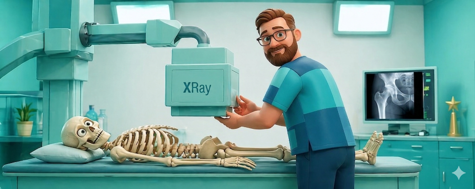
AP Hip
The AP Hip is the fundamental projection for evaluating the proximal femur and the hip joint. It is the primary tool for diagnosing hip fractures, particularly in the elderly, and assessing joint degeneration. Because the hip is a deep joint, the “Richie Standard” focuses on technical precision and a specific internal rotation to ensure the femoral neck is seen in its full, diagnostic profile.
The Goal
To demonstrate the acetabulum, femoral head, and femoral neck in a true anatomical profile, ensuring the neck is not foreshortened and the greater trochanter is profiled laterally.
Patient Positioning
- The Setup: Place the patient supine on the table with their arms comfortably at their sides or across their chest.
- The Rotation: Internally rotate the entire leg and foot 15–20 degrees. This movement overcomes the natural anteversion of the femur and places the femoral neck parallel to the image receptor.
- The Centering: Direct the Central Ray (CR) perpendicular to the IR, centered to the mid-femoral neck.
- Pro-Tip: If the patient is in extreme pain or a fracture is obvious, do not attempt internal rotation. Perform the image exactly as they lie and document the trauma status for the radiologist.
Technical Factors
- Central Ray (CR): Perpendicular to the IR, centered 1–2 inches distal to the midpoint of a line connecting the ASIS and the pubic symphysis.
- SID: 40″.
- Collimation: Include the ilium, acetabulum, and the proximal third of the femur. Ensure your light field extends to the lateral soft tissue margin.
Image Evaluation Criteria (The “Richie” Checklist)
A professional AP Hip image must pass these checks:
- Femoral Neck in Profile: The femoral neck should be seen in its full length without any foreshortening.
- Greater Trochanter: You should see the greater trochanter clearly profiled on the lateral aspect of the femur.
- Lesser Trochanter Location: The lesser trochanter should be nearly invisible, as it should be superimposed by the femoral neck.
- Joint Space Visualization: The hip joint space (acetabulum and femoral head) should be clearly visualized without rotation of the pelvis.
- Marker Visibility: Your physical R/L marker must be clear and positioned in the lateral collimation field, away from the bony anatomy.
Why the AP Hip Matters
This view is the diagnostic anchor for identifying Hip Fractures. Without the specific 15–20 degree internal rotation, the femoral neck rotates posteriorly and appears shortened on the image, which can hide subtle non-displaced fractures. By profiling the neck correctly, you provide the orthopedic surgeon with the accurate measurements needed for surgical hardware or joint replacement planning.
Richie’s Pro-Tips for the AP Hip
Physical Markers Only: Always place your physical lead marker on the IR at the time of exposure to ensure the side is correctly documented for the radiologist.
The “Tape” Trick: If a patient is unable to hold the internal rotation due to muscle weakness, use a long strip of medical tape across the toes or ankles to gently hold the position. It’s better than having a hand in the field!
Center of the Crease: If you’re having trouble palpating the ASIS on a larger patient, center the CR in the middle of the “inguinal crease.” This usually aligns perfectly with the femoral neck.
Expiration Breathing: Always have the patient hold their breath on expiration. This thins the abdominal density and provides a sharper, higher-contrast image of the deep hip structures.